Cephalic Vein Blood Draw Dog
This administration corresponds to a bolus injection at the cephalic vein. The parameters set in the model were m = 3, /r0 = 50 cm, Aq = 3 cm2, and b = 105. The estimated model parameters were ... [Pg.199]
Male or female inbred Beagle or Labrador-Harrier dogs weighing between 15 and 25 kg are used. They are anesthetized with a bolus injection of 35 40 mg/kg pentobarbital, and continued with an infusion of 4-6mg/kg/h. A catheter is placed into the cephalic vein for intravenous injections. Another catheter is placed into the duodenum for enteral administration. Respiration is maintained with room air through... [Pg.89]
FIGURE 45-1. The predominant types of vascular access for chronic dialysis patients are (A) the arteriovenous fistula and (B) the synthetic arteriovenous forearm graft. The first primary arteriovenous fistula is usually created by the surgical anastomosis of the cephalic vein with the radial artery. The flow of blood from the higher-pressure arterial system results in hypertrophy of the vein. The most common AV graft is between the brachial artery and the basilic or... [Pg.854]
Throat swabs, blood withdrawals, nasal washes and saliva samples are isolated on the indicated (X) days. Blood specimens (3 mL) are taken by venipuncture from the cephalic vein. Nasal washes are collected by instilling 1.5 mL of sterile phosphate-buffered saline (PBS) into the nostril and immediately aspirating wash fluid and secretions with a sterile S5uinge. Saliva specimens are collected by aspiration from the cheek pouch. Prior to throat swabbing, clinical scoring of tonsillitis and pharyngeal erythema severity is performed by a trained veterinarian who is blinded to the two animal groups. [Pg.260]
Take 3-mL blood samples from each of the anesthetized animals by venipuncture in the cephalic vein. [Pg.262]
A thorough understanding of the venous anatomic structures of the head, neck, and upper extremities are imperative for safe venous access (Fig. 4.2) (41). The precise location and orientation of the internal jugular, innominate, subclavian, and cephalic veins are important for safe venous access (42). Their anatomic relation to other structures is crucial in avoiding complications. The venous anatomy of interest from a cardiac pacing and ICD point of view starts peripherally with the axillary vein (43). [Pg.122]
Trapezius Clavicle Subclavlus Cephalic vein Pectorials major muscle (cut)... [Pg.122]
| Fig. 4.3 Anatomic relationship of the axillary vein to the pectoralis minor muscle. The pectoralis major has been removed. Note the cephalic vein draining directly into the axillary vein at approximately the first intercostal space. (From Belott PH. Unusual access sites for permanent cardiac pacing. In Barold SS, Mugica J, eds. Recent advances in cardiac pacing Goals for the 21st century. Armonk, NY Futura Publishing, 1997, with permission.)... |  |
| Table 4.9 Venous structures for pacemaker lead insertion. Cephalic vein Axillary vein Subclavian vein Internal jugular vein... | 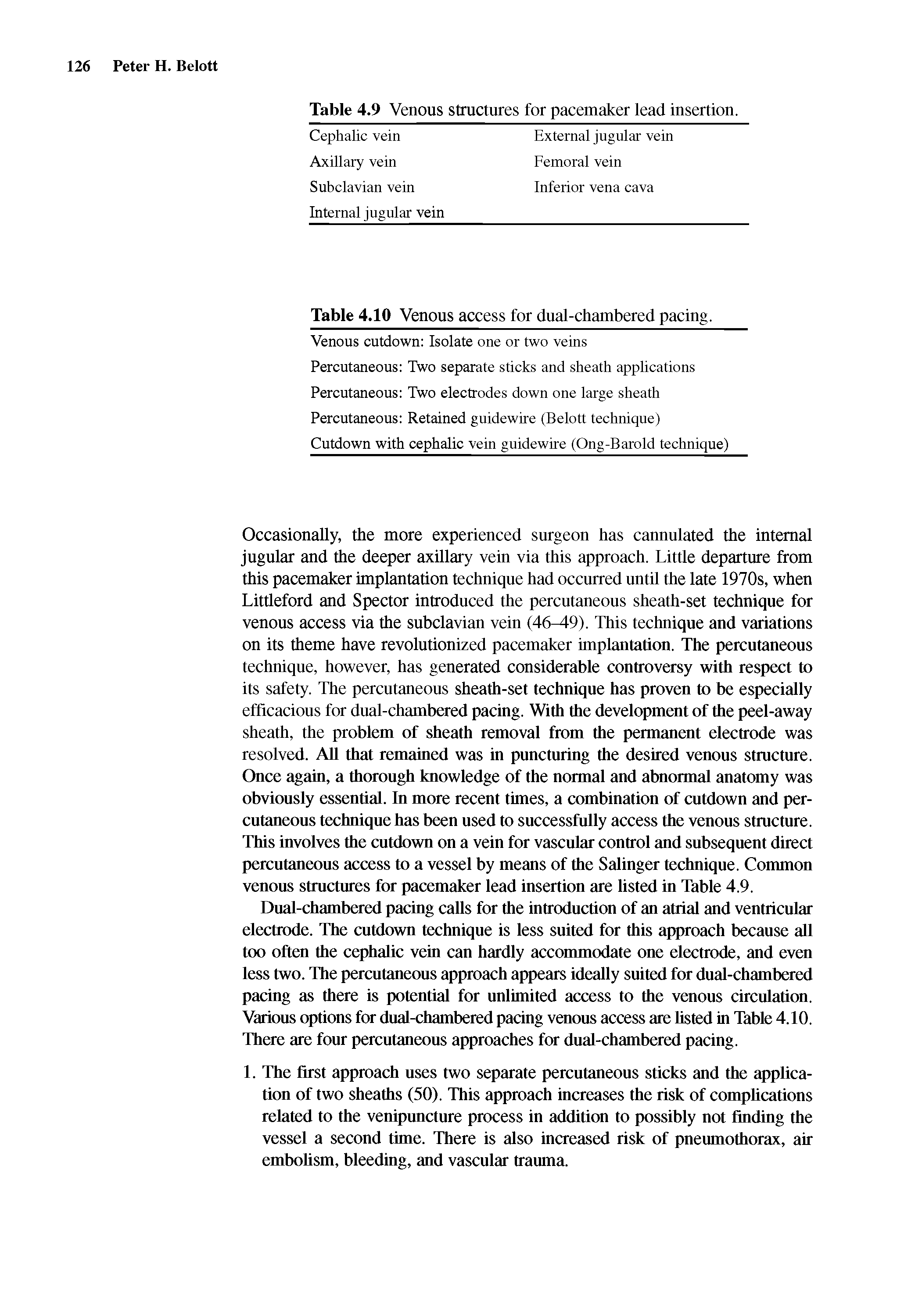 |
Dual-chambered pacing calls for the introduction of an atrial and ventricular electrode. The cutdown technique is less suited for this approach because all too often the cephalic vein can hardly acconunodate one electrode, and even less two. The percutaneous approach appears ideally suited for dual-chambered pacing as there is potential for unlimited access to the venous circulation. Various options for dual-chambered pacing venous access are listed in Table 4.10. There are four percutaneous approaches for dual-chambered pacing. [Pg.126]
Venous Cutdown of the Cephalic Vein Cephahc Venous Access... [Pg.127]
If the cephalic vein is too small, further dissection may be carried proximally. In rare instances, dissection will actually be carried to the deeper axillary vein. Once exposed, the cephalic vein is freed from its fibrous attachments and O silk ligatures are applied proximally and distally (Fig. 4.8). Once adequate venous control has been obtained, a horizontal venotomy is made with an iris scissor or a 11 scalpel blade (Fig. 4.9). The vein should be supported at all hmes with a smooth forceps. Using mosquito clamps, forceps, or vein pick, the venotomy is opened and the electrode(s) introduced (Fig. 4.10). Once venous access has been achieved, the electrodes are positioned in the appropriate chambers using standard techniques. [Pg.128]
| Fig. 4.8 Cephalic vein cutdown technique. The cephalic vein is isolated and tied off distally. (From Belott PH, Reynolds DW. Permanent pacemaker implantation. In EUenbogen KA, Kay N, WiUcoff BL, eds. Clinical cardiac pacing. Philadelphia WB Saunders, 1995, with permission.)... |  |
Ong LS, Barold S, Lederman M, et al. Cephalic vein guidewire technique for implantation of permanent pacemakers. Am Heart J 1987 114 753. [Pg.241]
Vamagy G, Velasquez R, Navarro D. New technique for cephalic vein approach in pacemaker implants. PACE 1995 18 1807a. [Pg.241]
Parsonnet V, Roelke M. The cephalic vein cutdown versus subclavian puncture for pacemaker/ICD lead implantation. PACE 1999 22 695. [Pg.246]
Venous access. In adnlt pacemaker practice, it is common to obtain a cut-down on the cephalic vein that will accommodate one or two leads. However, in children, becanse of the size of the vein, this is less likely. Still, the cephalic approach is preferable to the snbclavian approach, when available, as it completely avoids the complication of subclavian crush injury to the lead (39,40). Subclavian crnsh injnry resnlts from entrapment of the lead between the clavicle and the first rib, where it is subject to great stress with patient movement. [Pg.556]
Radiographic differentiation of the venous route used for lead placement, e.g., axillary, subclavian, and cephalic veins, may be difficult if not impossible. With implantation of the lead in an axillary or cephalic vein, the approach... [Pg.625]
When ultrasound guidance is used, a preliminary examination of the upper arm is performed to identify a suitable access vein. The basilic vein is the preferred site. The cephalic vein can be used,but is prone to spasm and has an acute angle as it joins the axillary vein. The veins are typically imaged in the transverse plane while the (echo-enhanced) needle is advanced (Fig. 6.4). The transducer is rocked cephalad and caudad keeping track of the needle tip. When this... [Pg.136]
Source: https://chempedia.info/info/cephalic_veins/
0 Response to "Cephalic Vein Blood Draw Dog"
Post a Comment